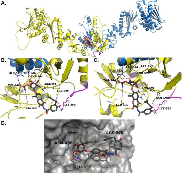
Figure 6. Modeled structure of the Novobiocin binding site in hHsp90α.
a) The Hsp90α homology model homodimer and the C-terminal binding site with NB (spheres) docked at the interface of two monomers (blue and yellow) b) close-up of NB (grey sticks) docked in Hsp90α homology model, the crosslinked fragment (lines) and predicted hydrogen bonds (dashes) are depicted in magenta and c) close-up of NB noviose (grey sticks) docked in Hsp90α homology model, predicted hydrogen bonds (dashes) are represented in magenta, d) surface representation of Hsp90α homology model CT binding site with NB (grey sticks) docked. Only one molecule of NB is shown to be bound to Hsp90 homodimer, an equivalent binding site contained on the other monomer has not been shown for clarity.

