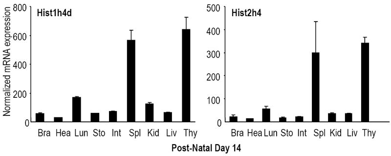
Figure 5. Proliferation-related tissue-specific expression of histone genes during post-natal development.

Expression levels of all mouse histone H4 genes were analyzed in selected tissues at multiple days during post-natal development (D1, D14 and D60) by qRT-PCR analysis normalized to Hprt1 (see Supplementary Figs. S2 and S3). The two graphs show representative results for two highly expressed histone H4 genes located in two different histone gene clusters (left, Hist1h4d; right, Hist2h4h) obtained for post-natal Day 14. Histone H4 genes are detectable in all tissues examined, including brain (Bra), heart (Hea), lung (Lun), stomach (Sto), intestine (Int), kidney (Kid), liver (Liv) and thymus (Thy).
