
Figure 6.
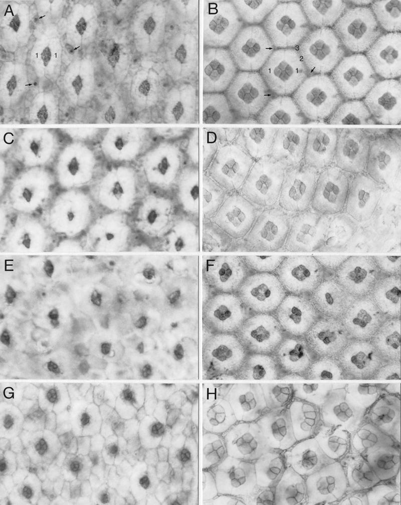
Abnormal development of cone and pigment cells in spa mutants. Early (24 hr APF at 25°C; A,C,E,G) and mid-pupal (45 hr APF at 25°C; B,D,F,H) eye discs of wild-type (A,B), homozygous spa1 (C,D), spaA/+ (E,F), and homozygous spapol (G,H) flies were stained with cobalt sulfide to visualize their cone and pigment cells at the apical surface of the retina. Examples of primary (1), secondary (2), and tertiary (3) pigment cells and of bristle cells (arrows) are marked in wild-type discs. Discs are shown with their anterior to the left and dorsal side up.