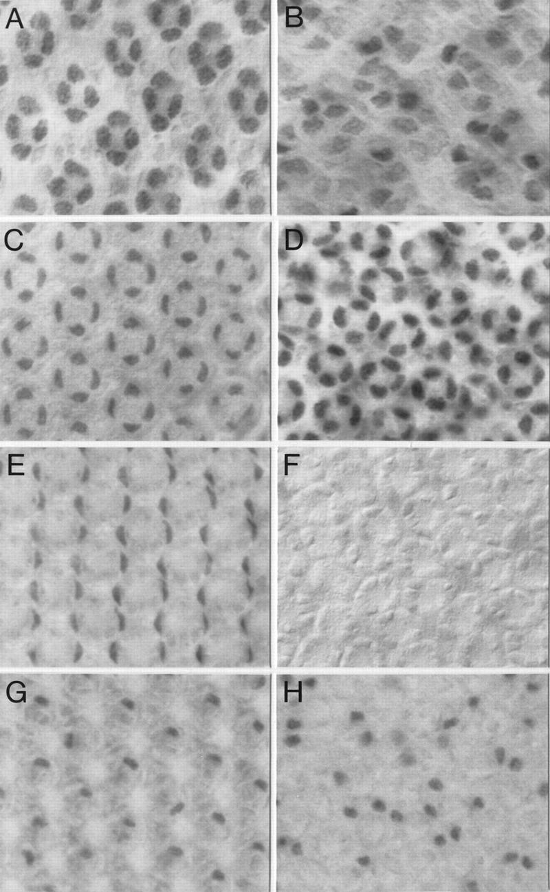
Figure 7.

Loss of Spa function affects expression of Cut and Bar proteins in cone and primary pigment cells of the eye disc. Cut expression in cone cells of a spapol (B), as compared to a wild-type (A), early pupal eye disc (24 hr APF at 25°C) is reduced. However, Cut expression in cone cells of a spapol mid-pupal eye disc (45 hr APF at 25°C; D) recovers and increases above the level observed in a wild-type mid-pupal eye disc (C). Bar expression in primary pigment cells of a wild-type (E) was also compared to that of a spapol (F) mid-pupal eye disc. Unlike the loss of its expression in primary pigment cells, Bar protein levels appear unaffected in bristle cells of a spapol (H) when compared to that of a wild-type mid-pupal eye disc (G). Note that the S12 anti-BarH1 antiserum used recognizes both BarH1 and BarH2 proteins (Higashijima et al. 1992b).
