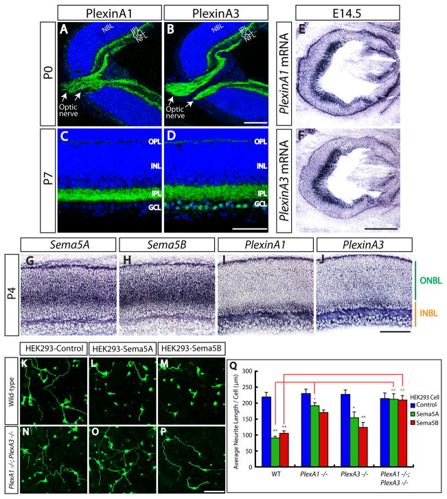
Figure 6. Sema5A/Sema5B and PlexinA1/PlexinA3 Exhibit Complementary mRNA Expression In the Developing Postnatal Retina, and PlexinA1 and PlexinA3 Mediate Inhibitory Responses of Retinal Neurons to Sema5A and Sema5B in vitro.
(A–D) Wild-type retina sections from postnatal P0 (A and B) and P7 (C and D) mice were immunostained with antibodies against PlexA1 (A and C) and PlexA3 (B and D). Both PlexA1 and PlexA3 are present in the nerve fiber layer (NFL) and the optic nerve (white arrows in A and B) at P0, revealing that PlexA1 and PlexA3 are expressed in RGCs. At P7, both PlexA1 and PlexA3 are present throughout the entire IPL. NBL, neuroblast layer.
(E and F) In situ hybridization for PlexA1 (E) or PlexA3 (F) on adjacent E14.5 embryonic retina sections. Both PlexA1 and PlexA3 are expressed in the INBL.
(G–J) In situ hybridization for Sema5A (G), Sema5B (H), PlexA1 (I), or PlexA3 (J) on P4 retina sections. Strong expression of Sema5A and Sema5B mRNA was detected in the ONBL, whereas strong PlexA1 and PlexA3 mRNA expression was restricted to the INBL.
(K–P) In vitro neurite outgrowth assay using dissociated retinal neurons from E14.5 wild-type (K–M) and PlexA1−/−; PlexA3−/− (N–P) embryos, cultured for 48hr on confluent monolayers of HEK293 cell lines expressing Sema5A, Sema5B, or transfected with a control expression vector. Wild-type retinal neurons cultured on Sema5A- or Sema5B-expressing cells exhibit reduced neurite extension (average neurite length per neuron) as compared to cells transfected with a control vector (K–M). However, inhibitory responses to Sema5A and Sema5B are not observed in PlexA1−/−; PlexA3−/− retinal neurons (N–P).
(Q) Quantification of average neurite length per neuron from in vitro neurite outgrowth assays (K–P), utilizing retinal neurons from E14.5 PlexA1−/−, PlexA3−/−, and PlexA1−/−; PlexA3−/− mutant embryos. Both Sema5A and Sema5B significantly inhibit neurite outgrowth from wild-type retinal neurons in vitro (see Figure 3K–3N). However, neurite outgrowth inhibition by Sema5A or Sema5B was abolished when PlexA1−/−; PlexA3−/− retinal neurons were used in this assay, and it was significantly attenuated for PlexA1−/− single mutant embryonic retinal neurons cultured on Sema5A-expressing HEK293 cells. The average neurite length per neuron: For PlexA1−/− embryos −231.1 ± 17.0 μm for control (n=56), 192.9 ± 15.0 μm for Sema5A (n=59), and 171.0 ± 12.4 μm for Sema5B (n=62); For PlexA3−/− embryos −227.3 ± 15.0 μm for control (n=58), 154.7 ± 14.2 μm for Sema5A (n=60), and 124.0 ± 10.5 μm for Sema5B (n=53); For PlexA1−/−; PlexA3−/− embryos −199.7 ± 16.2 μm for control (n=49), 195.2 ± 19.3 μm for Sema5A (n=57), and 196.1 ± 13.7 μm for Sema5B (n=46). Error bars are SEM (n=3 independent experiments). Black * indicates statistical significance of neurite outgrowth on ligand-expressing HEK293 cells as compared to control HEK293 cells within each genotype. Red * indicates statistical significance under the same treatment between wild-type and other retinal neuron genotypes. * and ** show P < 0.05 and P < 0.01, respectively, by multi-factorial ANOVA followed by Tukey’s HSD test.
Scale bars: 100 μm in B for A and B, 50 μm in D for C and D, 500 μm in F for E and F, 100 μm in J for G–J, and 100 μm in P for K–P.

