
Figure 4.
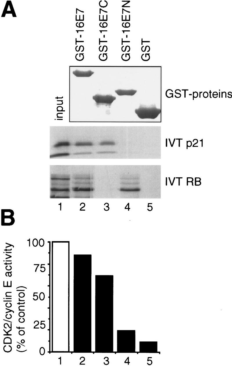
Analysis of binding sites in HPV-16 E7 proteins. (A) In vitro binding reactions with GST–16E7 mutants (1.5 μg) immobilized on glutathione–Sepharose and IVT p21 or IVT RB were performed, and the resulting protein complexes were visualized by Coomassie staining (top) and fluorography (bottom). Lane 1 represents 10% (IVT p21) or 20% (IVT RB) of the input. (B) Purified recombinant His–cyclin E/CDK2 complexes were coincubated with buffer (□) or coincubated with His–p21 (10 ng, ▪) alone or with His–p21 preincubated with GST fusion proteins (300 ng; as indicated in A, lanes 2–5), and kinase assays were performed. The quantitation from PhorphorImager analysis is shown.