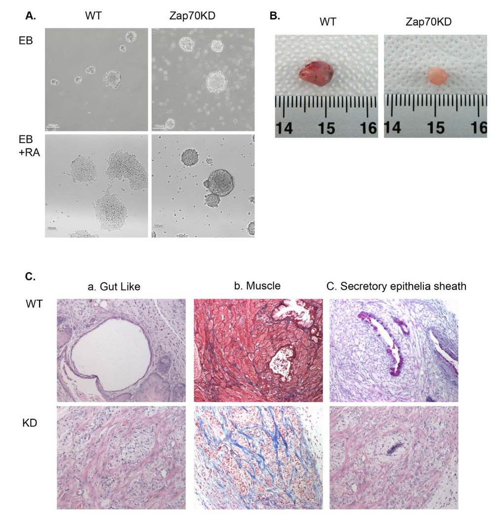
Figure 4. Defective differentiation capacity of Zap70KD.
(A) Embryonic bodies (EBs) were formed from either wild type or Zap70KD ES cells after 4 days in culture and differentiation was induced by the addition of retinoic acid. Cell morphology was examined 3 days after differentiation. Scale bar is 100 mm. (B) Morphologies of mESC teratomas obtained from NOD/SCID mice injected with either wild type or Zap70KD are shown. Scale bar, 1cm. (C) Histology staining of mESC induced teratomas stained with H/E, Masson’s trichrome, and Alcian Blue are shown. Gut-lie, muscle, secretory epithelia sheath components were observed in wild type mESC.

