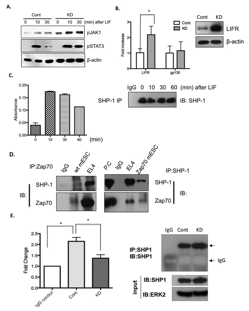
Figure 5. LIFR expression and SHP-1 phosphatase activity are two mechanisms for Zap70 to regulate Stat3 signals.
(A) mESCs LIF-starved for 24 h were stimulated with LIF for the indicated time and then active phosphorylation of Jak1 (top panel) and Stat3 (middle panel) were determined by immunoblotting. β-actin was used as protein loading control (bottom panel). (B) LIFR and gp130 mRNA level were determined by qRT-PCR and graphically presented. *, p<0.05. LIFR protein expression was shown by immunoblotting analysis. β-actin was used as protein loading control (right panel). (C) mESCs LIF-starved for 24 h were treated with LIF for the indicated time and then harvested. Left panel: SHP-1 enzymatic activity was measured, followed by SHP-1 immunoprecipitation. Right panel: immunoprecipitated SHP-1 levels were compared by immunoblotting (IB) for SHP-1 followed by SHP-1 IP. (D) Co-immunoprecipitation analysis of the interaction of endogenous Zap70 (left panel) or ectopically expressed Zap70 (right panel) with SHP-1. Interaction between Zap70 and SHP-1 in EL4 was used as a positive control. Non-specific IgGs were used as negative control. P.C, positive control from EL4 whole cell lysate. (E) Left panel: SHP-1 enzymatic activity followed by immunoprecipitation. *, p < 0.05. Right panel: Immunoprecipitated (IP) SHP-1 level, determined by immunoblotting (IB) for SHP-1 followed by SHP-1 IP. SHP-1 and ERK2 immunoblotting of whole cell lysate (input) from control mESC (Cont) and Zap70KD (KD) were used as control of equal protein concentration for the IP and phosphatase activity assays.

