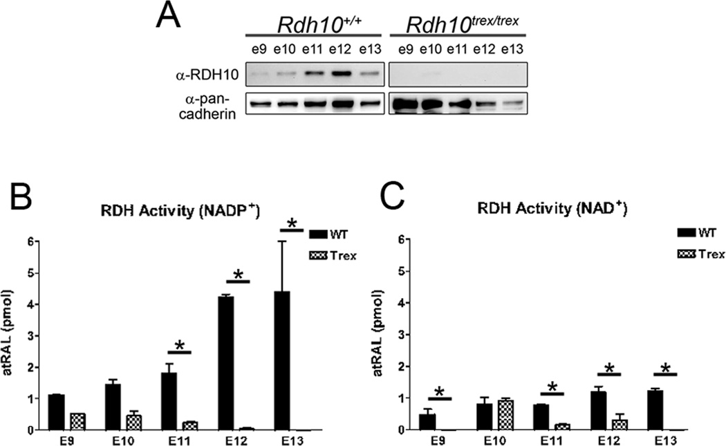
Figure 3. Rdh10trex embryos have a significant reduction in total RDH activity.
(A) Whole embryo membrane fractions were prepared for RDH activity assays and analyzed by Western blotting (20 µg of protein per lane) using an anti-RDH10 antibody and an anti-pan-cadherin antibody as a loading control. (B and C) RDH activity was measured with either (B) NADP+ or (C) NAD+ cofactor. The bar graphs represent the mean +/− s.d. from at least three independent experiments (*p<0.05 by two-way ANOVA).

