Figure 6.
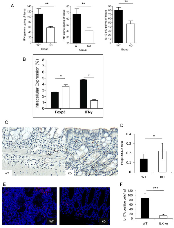
Epithelial ILK regulates tissue expression of inflammatory cytokines. A. Interferon gamma, tumor necrosis factor alpha and interleukin-12p40 cytokine levels were determined in colonic homogenates from 6 ILK-knockout animals and 6 wild-type controls (**p < 0.01). B. Lymphocytes were obtained from mesenteric lymph nodes of wild-type and ILK-ko mice. Intracellular staining for FoxP3 and IFNγ was performed as described in materials and methods. After stimulation with PMA (25 ng/ml) and ionomycin (1 mg/ml) for 6 h, cells were fixed and permeabilized. Then they were stained with the indicated antibodies and read on a BD FACS Canto. The data are from 6 ILK-ko and 6 wild-type mice (*p < 0.05). C. Tissue sections were obtained from control and ILK-ko mice at the end of 3 rounds of DSS treatment, and processed for immunohistochemistry. Using anti-CD3 and anti-FoxP3 antibodies, the number of positively staining cells were counted in 3 fields from 6 separate animals, in each group. The ratios obtained are shown in D (*p < 0.05). E. IL-17A staining was performed using immunofluorescence as described in methods for tissue sections from the same sets of mice as in C. The red staining cells are clearly observed to be more numerous in the control samples, and the data is graphically represented in F.

