
Figure 1.
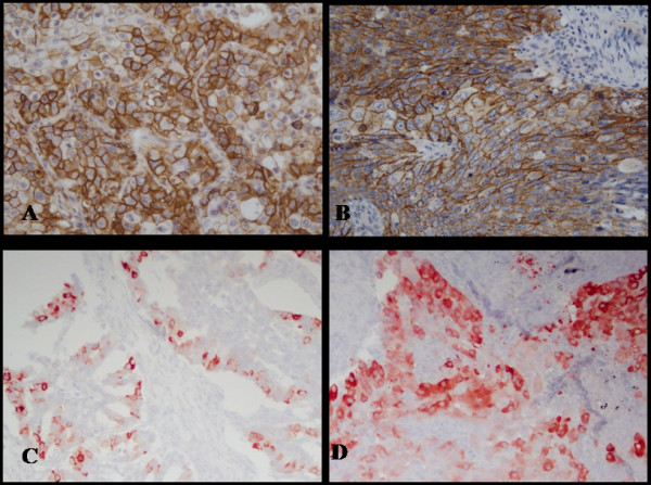
Microscopic views of positively stained immunohistochemistry sections in non-small cell lung cancer. Immunoreactivity for CD44s was detected predominantly in the membrane of tumor cells. Typical examples of CD44s immunopositivity in AC (×400; A) and SCC (×400; B) histological subtypes. Immunoreactivity of COX-2 was detected predominantly in the cytoplasm of tumor cells. COX-2 immunopositivity in AC (×400; C) and SCC (×400; D) histological subtypes.