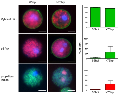
Figure 1. The membrane of freshly formed detached cells is intact and retains phosphatidylserine asymmetry.
HepG2 cells were infected with fluorescent P. berghei parasites and detached cells were collected at either 65 or >75 hours of infection. They were then stained with the nuclear dye Hoechst 33342 (blue) and several additional live stains to characterize the nature of the surrounding membrane. The existence of a membrane in general was shown by staining with Vybrant DiO (green) in cells infected with P. berghei-mCherry (red, top panel). The presence of phosphatidylserine in the outer membrane leaflet, an indicator for ongoing cell death, was tested using pSIVA (green) in cells infected with P. berghei-mCherry (red, middle panel). Propidium iodide (red) is a membrane-impermeable nuclear stain and served to check the integrity of the membrane of P. berghei-GFP (green)-infected detached cells (lower panel). Quantification of the phenotypes (mean + SEM of three independent experiments) clearly shows that the membrane of freshly formed detached cells was intact and phosphatidylserine-negative while the membrane of older detached cells slowly lost phosphatidylserine asymmetry and integrity. Total n for 65hpi - Vybrant DiO: 712, pSIVA: 1483, PI: 1291. Total n for 75hpi - Vybrant DiO: 899, pSIVA: 874, PI: 1564. Bars = 10 µm; WF (wide-field).

