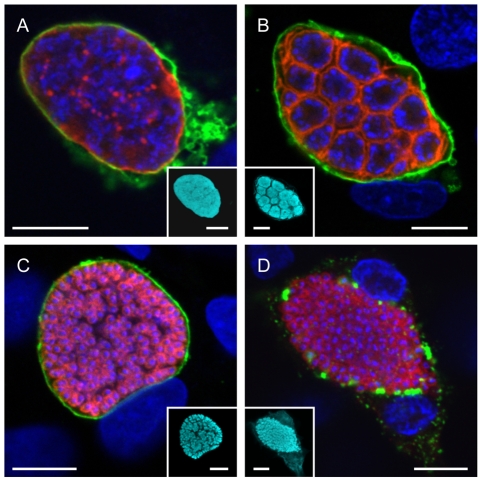
Figure 2. The parasite membrane becomes the merozoite membrane during the late liver phase.
HepG2 cells were infected with P. berghei parasites and fixed at different time points after infection. They were then stained with an anti-MSP1 antibody to visualize the PM (red) and anti-Exp1 antibody to label the PVM (green). The parasite cytoplasm was labeled using a transgenically expressed protein (cyan) and the nuclei were stained with DAPI (blue). While the PM surrounded the parasite as a whole during the late schizont stage (A), it began to invaginate around groups of nuclei during cytomere formation (B). It eventually surrounded individual merozoites both before (C) and after (D) PVM breakdown. Representative images are shown. Bars = 10 µm; CPS (confocal point scanning).

