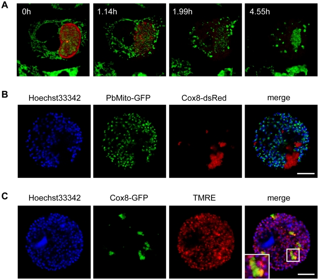
Figure 6. Host cell mitochondria disintegrate shortly after PVM breakdown.
A HepG2 cells were infected with P. berghei-Exp1-mCherry and stained with MitoTracker GreenFM at 62 hpi. Stills from a representative movie (Video S6) show that host cell mitochondria drew together and disintegrated. n = 17 from three independent experiments, bar = 10 µm, CLS. B pDsRed1-N1-Cox8-transfected HepG2 cells were infected with Pb cGFPMITO parasites to visualize parasite mitochondria (green). Detached cells were collected at 68 hpi and stained with Hoechst 33342 (blue). The overlay shows that the mitochondria remnants were of host cell origin. Representative images are shown. Bars = 10 µm, CPS. C pEGFP-N1-Cox8-transfected HepG2 cells were infected with P. berghei parasites. Detached cells were collected at 68 hpi and stained with Hoechst 33342 (blue) and TMRE (red). Representative images show that most of the host cell mitochondria remnants (green) had no TMRE staining, indicating a loss of membrane potential. An enlarged part of the merged image (indicated by a white box) is shown in the inset. Bars = 10 µm, CPS.

