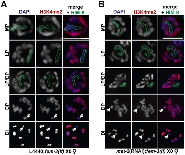
Figure 2. Ectopic X-specific H3K4me2 accumulation in late pachytene X0 germ lines depleted for met-2.
Immunolocalization of H3K4me2 (red) counterstained with DAPI (blue) in fem-3(lf) X0 germ lines fed (A) empty L4440 vector (left) or (B) met-2 dsRNA (right). Green outline indicates the X chromosome in MP, LP, and LP/DP, as determined by HIM-8 staining (green). White arrows indicate the X chromosome in DP and DI. Mid-pachytene (MP); Late pachytene (LP); diplotene (DP); diakinesis (DI). Scale bar = 5 µm. (See also Figure S4).

