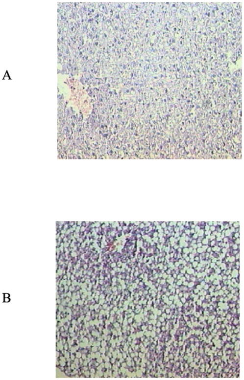
Figure 1. Histological analysis of liver specimens stained with haematoxylin and eosin (H&E) from controls (A) and rats fed the MCD diet (B).
Liver of rats fed the MCD diet shows steatohepatitis characterized by panacinar steatosis predominantly as macrovescicular fat, together with lobular inflammation (H&E ×100).

