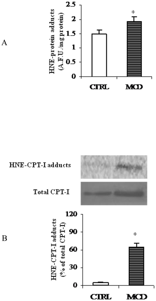
Figure 7. HNE-protein adducts in liver mitochondria.
(A) Levels of HNE-protein adducts in liver mitochondria from methionine-choline deficient (MCD) diet and control rats (CTRL) were measured by fluorimetric analysis and evaluated in terms of arbitrary fluorescent units (A.F.U.) at 355 nm excitation and 460 nm emission. Data are expressed as means ± SD of five experiments for each group. (B) Western blot analysis was performed to reveal HNE-CPT-I adducts from CTRL and MCD rats. Signals were quantified by densitometric analysis and expressed as % of the total CPT-I protein content measured in mitochondria from CTRL and MCD rats. Statistical differences were assessed using unpaired t-test assuming variance homogeneity. *Significantly different from the control.

