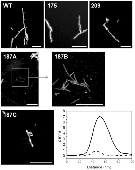
Figure 3. AFM reveals the presence of thin fibrils in addition to mature fibrils in conversion reactions of T187I.
Conversion reactions of wild-type (WT), V175I, V209I (upper row) and T187I (middle, bottom row) were observed under the atomic force microscope. In all samples fibrils with approximate height around 7 nm were observed. Cross-section of such fibril from sample T187I is shown (bottom row right, full line). In the T187I sample in addition to such fibrils, fibrils with less than 1 nm in height were also present (middle row). Image 187B shows a close-up of selected part in image 187A. Cross-section of thin fibril is shown in the bottom row (bottom row right, broken line). Bar represents 500 nm. Arrows in images 187B and 187C indicate positions where cross-sections were taken. Images have not been corrected for the width and shape of the AFM tip.

