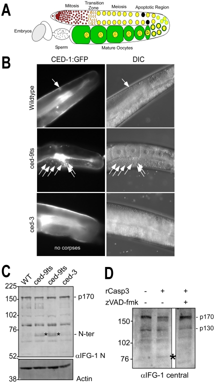
Figure 1. IFG-1 is cleaved during C. elegans apoptosis in vivo and by human caspase-3.
(A) Diagram depicting one lobe of the adult gonad. Germ cells begin as a stem cell population of mitotically dividing cells in a common cytoplasm that transitions into a meiotic program. As oocytes mature they become fully cellularized and accumulate cytoplasmic components and condensed bivalent chromosomes prior to fertilization. The region of naturally occurring apoptotic cell death is indicated. (B) Fluorescence images depicting germ cell apoptotic events induced by loss of ced-9 function. One lobe of the gonad from adult wild type, temperature-sensitive ced-9ts (n1653), and ced-3 (n2452) strains expressing the apoptotic marker CED-1::GFP are shown. GFP-positive apoptotic corpses are designated by white arrows. Overlapping DIC images to the right demonstrate normal gonad and oocyte morphologies. In the wild type and ced-3 panels, the path of the intestine has obscured fully grown oocytes in the proximal arm, but younger oocyte nuclei are visible in the distal arm prior to the bend, in the region where germ cell apoptosis occurs. (C) Western blot of IFG-1 p170 cleavage in vivo in ced-9(ts) adult worms. Worms treated 48h at 25°C were homogenized and the extract analyzed by 8% SDS-PAGE. Two independent lines of ced-9ts (n1653) worms were analyzed. Immunoblotting was performed using IFG-1 N-terminal and actin antibodies. N-terminal cleavage products are indicated (*). (D) Immunoblot analysis demonstrating ex vivo cleavage of IFG-1 p170 from total wild type C. elegans lysate by purified recombinant human caspase-3 (Sigma). The blot was probed with an IFG-1 central domain antibody that detects both p170 and p130 isoforms. The migration of cleavage products is indicated (*).

