Figure 3.

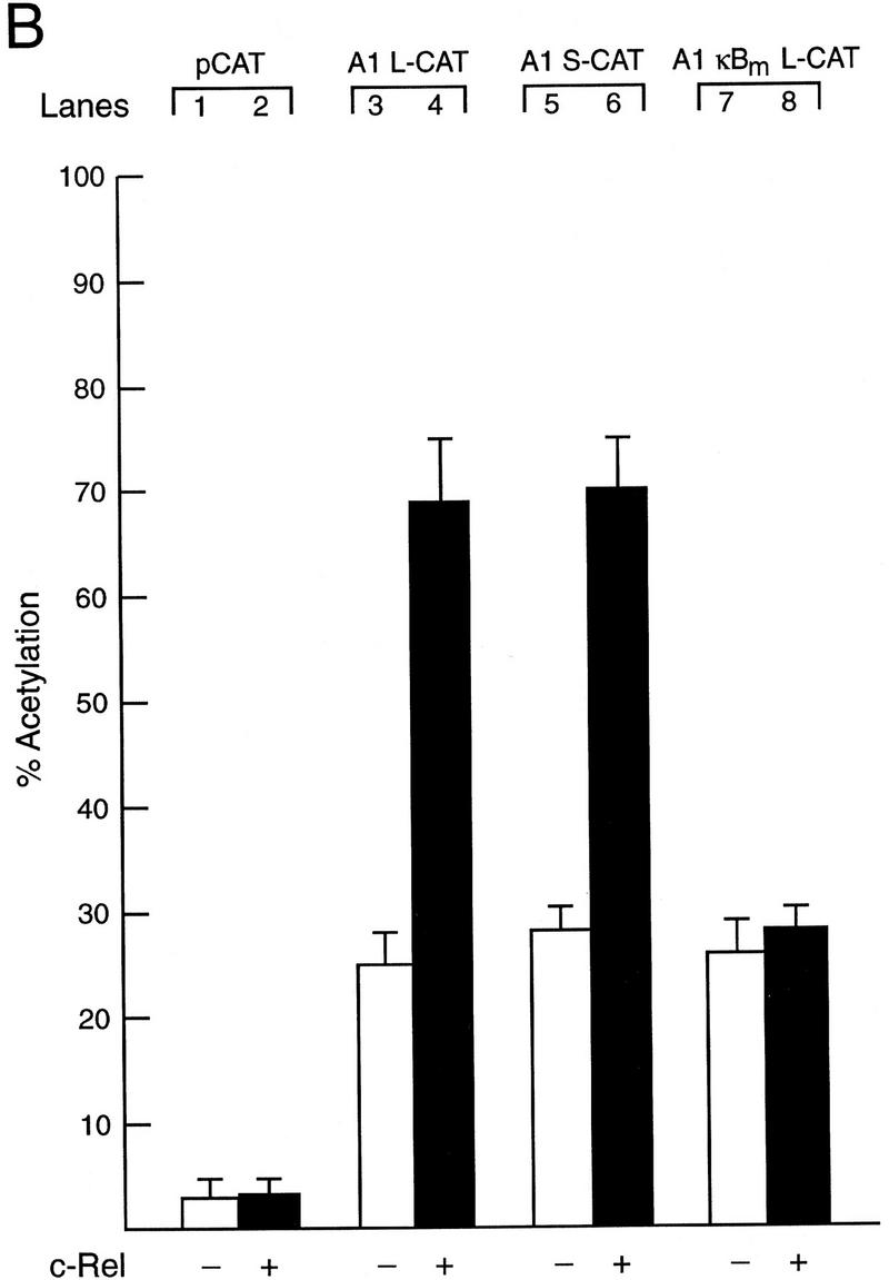
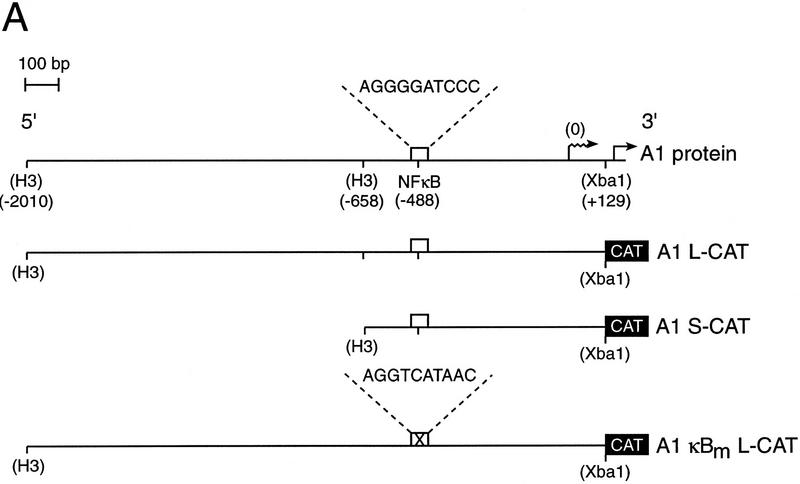
Functional analysis of the murine A1 promoter. (A) Schematic diagram of the A1 5′ flanking region and the CAT reporter plasmids. The numbers in parenthesis indicate the positions of restriction enzyme sites introduced into the murine A1 5′ flanking region by in vitro mutagenesis according to the numbering of the sequence outlined in Fig. 2. The open box represents the putative NF-κB-binding site (5′-AGGGGATCCC-3′: −488 to −479), whereas the corresponding symbol with a cross represents the mutated motif (5′-AGGTCATAAC-3′). The wavy arrow denotes the transcription initiation site and the straight arrow denotes the murine A1 initiation codon. The CAT gene is depicted as a closed box. Plasmid nomencature is indicated to the right of each construct. (B) Mutation of the NF-κB-binding motif abolishes Rel-dependent transcription in T cells. Jurkat cells were transfected transiently with 2 μg of the reporter plasmids pCAT (lanes 1,2), A1 L-CAT (lanes 3,4), A1 S-CAT (lanes 5,6) or A1κBm L-CAT (lanes 7,8) plus 10 μg of the expression plasmid DAMP56 containing no insert (lanes 1,3,5,7) or the c-rel expression plasmid pDAMP56c-rel (lanes 2,4,6,8). Chloramphenicol acetylation for transfections with the pDAMP56 or pDAMP56c-rel are indicated by open and closed bars, respectively. These results represent the mean percentage of chloramphenicol acetylation ± s.d. obtained from five separate sets of transient transfections.

