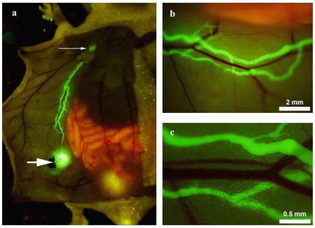
Fig. 1.
In vivo fluorescence imaging of the ventral skin lymphatic system in a skin flap mouse model. AlexaFluor-conjugated anti-LYVE-1 antibody was injected into the inguinal lymph node (A, large arrow); under fluorescence imaging, the inguinal lymph node, afferent lymphatics, and axillary lymph node (A, small arrow) display a strong, durable fluorescent signal that did not stain adjacent blood vessels (B,C).
From ref. 15.

