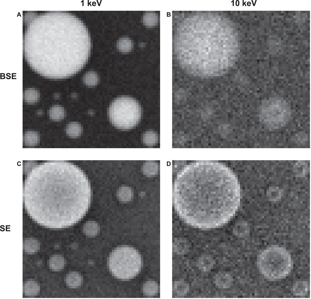
Figure 5.
Simulated images of tin balls on a carbon substrate. A, B: backscattered electron images. C, D: secondary electron images. The incident energy of 1 keV (A, C) and 10 keV (B, D). The tin ball diameters are 20, 10, 5, and 2 nm. The field of view is 40 nm with a pixel size of 0.5 nm. The nominal number of electrons for each scan point was 1,000.

