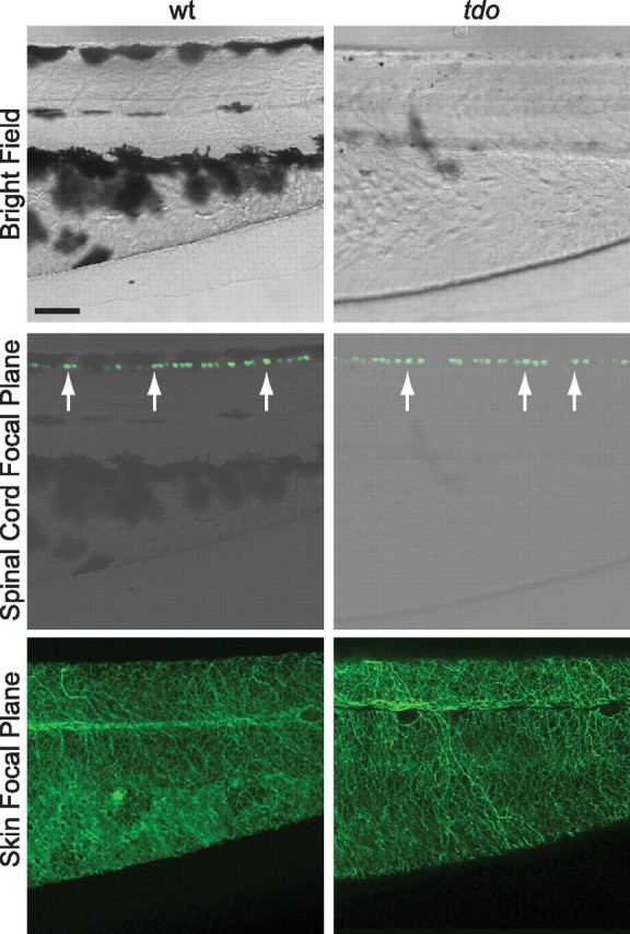
Figure 7.

The gross morphology of RBs appears normal in touchdown mutants. Top, A lateral bright field view of the trunk and tail regions from ssx-mini-ICP:eGFP transgenic wild-type and touchdown mutant larvae at 55 hpf. Rostral is to the left and dorsal is up. Scale bar, 50 μm. Middle, Confocal microscope view of the same regions displaying RB cell bodies (detected by using anti-GFP and a fluorescent secondary antibody) superimposed over the bright field image. The microscope was focused at the level of the RB cell bodies. Bottom, The same larvae viewed in a different focal plane that highlights RB peripheral neurites within the skin.
