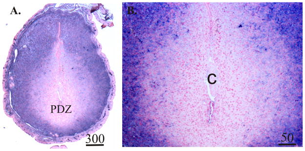
Fig. 3.
Representative photomicrographs of biotinylated-albumin histochemistry of sections prepared from Day 5.5 pregnant mice. Tissues were collected 1 hour after intravenous injection of biotinylated albumin. Values above scale bars represent microns. Sections from the uteri of mice that were not injected with biotinylated albumin showed no purple staining (not shown). PDZ, primary decidual zone of the endometrium; C, implanting conceptus.

