
Figure 2. Hypertrophy and heart failure develop when doxycycline is withheld from tTA/CnA* mice.
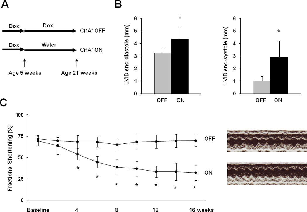
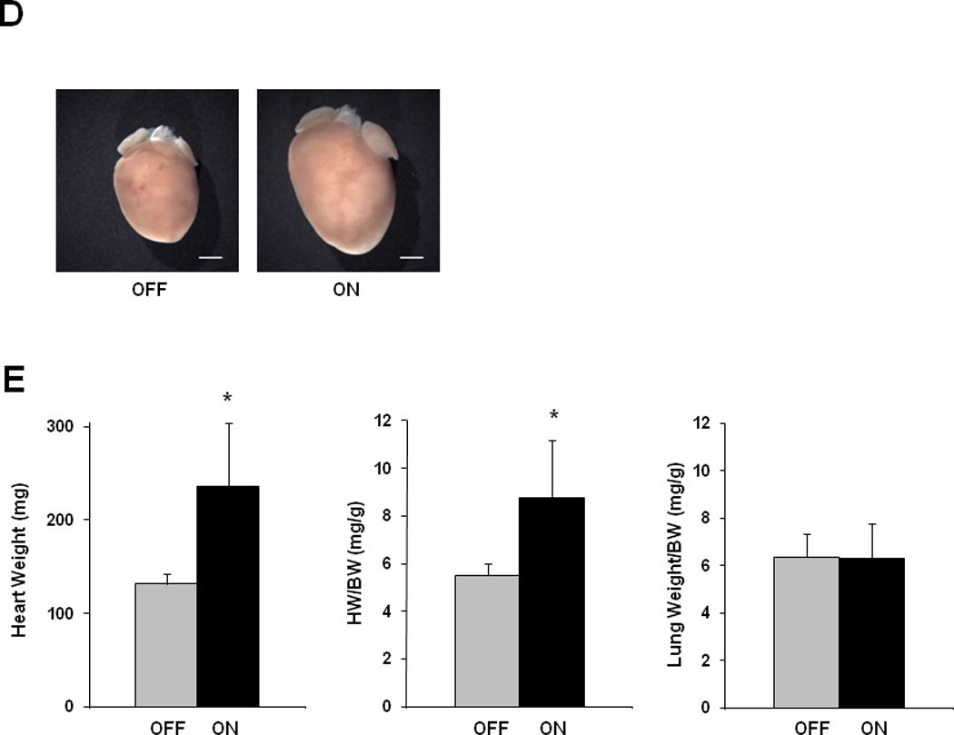
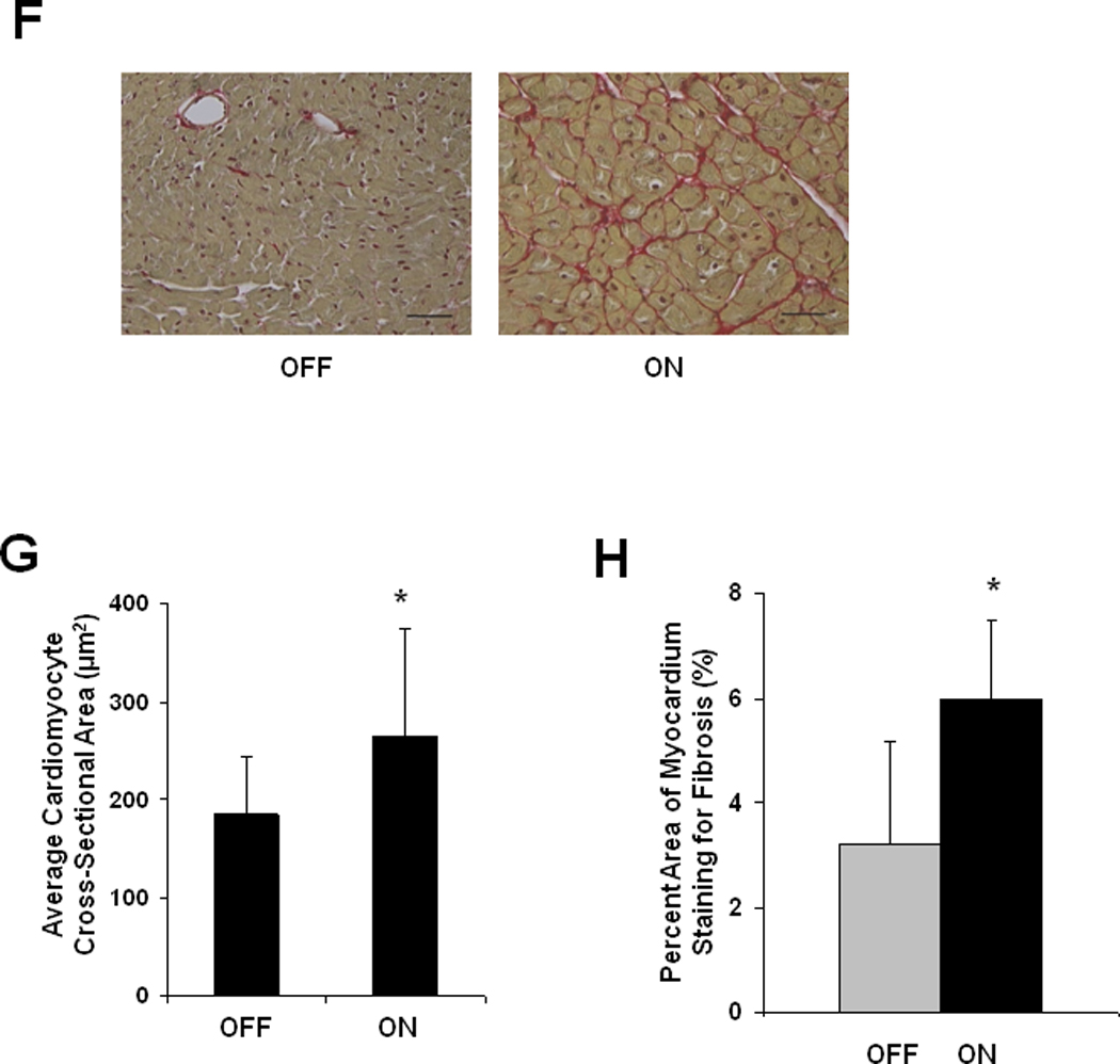
A: Experimental design. Double transgenic tTA/CnA* mice maintained on doxycycline (OFF) were used as controls. B: Left ventricular internal diameter (LVID) at end-diastole and end-systole after 16 weeks of calcineurin activation. C: Mean percent fractional shortening measured from M-mode tracings from mice in the OFF (n=10) and ON (n=14) groups over the course of the experiment. Representative M-mode echocardiographic tracings showing left ventricular mechanical activity at study completion. D: Representative images of hearts at study completion (scale bar = 800 µm). E: Mean absolute heart weight, heart weight normalized to body weight (HW/BW), and lung weight normalized to body weight for mice in the OFF (n=10) and ON (n=14) groups at study completion. F: Representative histological images of picrosirius red staining revealing cardiomyocyte size and fibrosis at study completion. Bar = 80 µm. G: Mean cardiomyocyte cross-sectional area measured from hearts in the OFF and ON groups (4 hearts per group; 60 cells measured per heart; bar = 1 SD). *p<0.01. H. Mean percent area of myocardium staining for fibrosis in the OFF and ON groups (6 images per heart, 6 hearts per group).