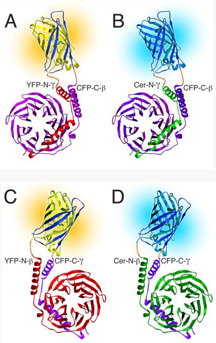
FIG. 1.
Models of fluorescent βγ complexes produced with multicolor BiFC. The split fluorescent protein at the top of each model is joined by linkers (orange) to the βγ dimer at the bottom. The CFP-C fragment (dark blue) is combined with either the YFP-N fragment (yellow) to produce yellow fluorescence or the Cer-N fragment (cyan) to produce cyan fluorescence. (A and B) YFP-N-γ and Cer-N-γ compete for CFP-C-β. β is magenta and γ is red (A) or green (B). (C and D) YFP-N-β and Cer-N-β compete for CFP-C-γ. γ is magenta and β is red (C) or green (D). The structures of the fluorescent protein fragments are adapted from the structure of GFP (16). The structures of β and γ are from the structure of an αt/αi1 chimera complexed with βtγt (17). (Reprinted from (18) with permission from Elsevier.)

