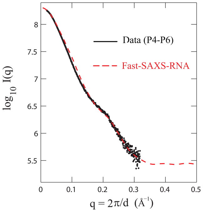
Figure 4.
Model validation of the Fast-SAXS-RNA method. The two-particle model with the hydration layer is validated using the experimental SAXS data and the crystal structure for the P4-P6 fragment of group I intron (P4-P6; PDB entry 1GID). Comparison of the experimental (black) and Fast-SAXS-RNA computed (red) SAXS profiles. The χ2 difference (Eq. (10)) between these two curves is 1.8 × 10⁻³.

