
Figure 6.
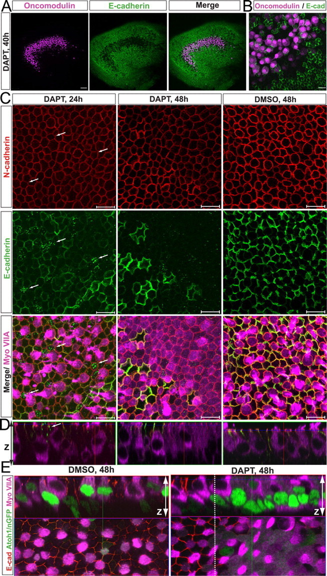
DAPT induces downregulation of E-cadherin and subsequent upregulation of myosin VIIA in striolar SCs. A, Image of a utricle treated with DAPT for 40 h and immunostained for oncomodulin (purple) and E-cadherin (green). Depletion of E-cadherin occurs specifically in the striolar region, which shows immunostaining for the type I hair cell marker oncomodulin. Scale bar, 50 μm. B, Higher-magnification image of the striolar region. Scale bar, 10 μm. C, Higher-magnification images of the striolar sensory epithelium. Arrows point to cells that show E-cadherin internalization. Myosin VIIA is upregulated after 48 h of DAPT treatment in cells of the striola. Scale bars, 10 μm. D, Images showing z-axis of sensory epithelium of utricles treated with vehicle or DAPT for 24 or 48 h. Arrow points to cells that show E-cadherin internalization. E, Images showing x–y-axis and z-axis of Atoh1/nGFP P2 utricles treated with vehicle (DMSO) or DAPT and immunostained for E-cadherin (red) and myosin VIIA (purple). The image of the DMSO control utricle was taken at the striolar/peristriolar boundary. In the image of the DAPT-treated utricle, the extrastriolar region is to the left of the white dashed line, and the striolar region is to the right.