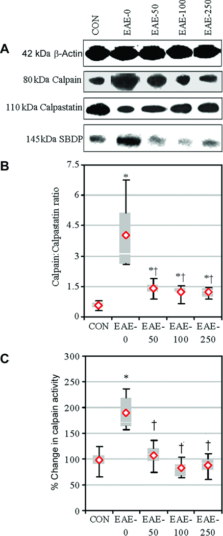
Fig. 3.
Calpeptin attenuated calpain expression and activity in acute EAE. Expression of calpain and calpastatin and activity of calpain were examined in tissues via Western blotting. (A) Representative blots showed calpain and calpastatin expression. Calpain activity was assessed using antibody against spectrin, which recognized the calpain-cleaved 145-kDa SBDP. (B) The mean calpain:calpastatin ratio compared with CON and (C) calpain activity as % change compared with CON-0 set at 100%. Results were presented as box-plot of inter-quartile data with the white line denoting the median expression. Diamonds represented mean protein expression (n = 4–5 per group, * P ≤ 0.05 compared with CON, and † P ≤ 0.05 for EAE-0 versus EAE-calpeptin).

