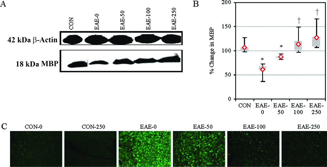
Fig. 5.
Calpeptin attenuated MBP degradation and axonal degeneration in acute EAE. (A) Representative blots and (B) average scanning densitometry presented as a % change in MBP compared with CON-0 set at 100%. Results were presented as box-plot of inter-quartile data with the white line denoting the median expression. Diamonds represented mean protein expression (n = 4 to 5 per group, * P ≤ 0.05 compared with CON, and † P ≤ 0.05 for EAE-0 versus EAE-calpeptin). (C) Representative images of axonal damage that was assessed by staining with SMI-311 antibody to detect de-NFP in the spinal cord tissues from CON and acute EAE Lewis rats after vehicle or calpeptin (50 – 250 µg/kg) therapy (n = 2 for CON groups, n = 4 to 5 for EAE groups, and magnification 200×).

