
Figure 4.
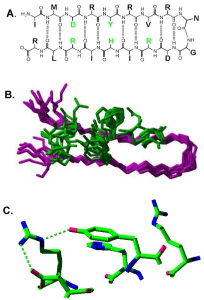
Structure of a beta hairpin peptide, IMDRYRVRNGDRIHIRLR, in which a tyrosine-histidine pi-pi interaction lowers the tyrosine redox potential and a PCET reaction occurs between tyrosine and histidine. (A) shows the primary sequence and some of the predicted hydrogen bonds and cross strand interactions; (B) is the overlap of the 20 lowest energy 2-D NMR structures, with only five amino acid side chains shown (see part A, green); and (C) is the redox active tyrosine and its immediate environment in the averaged, minimized NMR structure. Predicted hydrogen bonds are shown with dashed lines. Reprinted with permission from [112]. Copyright year 2007 American Chemical Society.