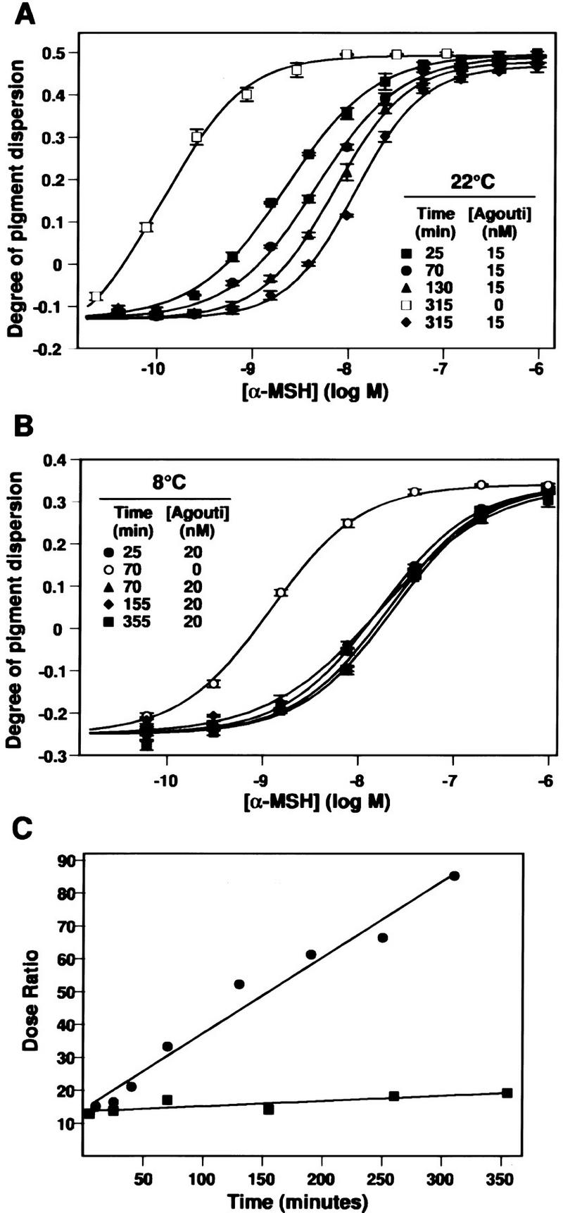
Figure 3.

Time and temperature dependence of Agouti protein action. (A) Melanophores were preincubated with 15 or 0 nm (buffer only) Agouti protein for the indicated length of time at 22°C, various concentrations of α-MSH were added for an additional 30 min, and the degree of pigment dispersion was determined as described in Materials and Methods. Preincubation in buffer does not alter the response to α-MSH, therefore, only one time point (315 min) is shown. Only four of the eight preincubation times in Agouti protein (25, 70, 130, and 315 min) are displayed. (B) Preincubation at 8°C. Same as in A except melanophores were kept at 8°C during preincubation with 20 nm Agouti protein, then incubated at 22°C following addition of α-MSH. Qualitatively similar results (not shown) are obtained if the entire experiment is carried out at 8°C. (C) Kinetics of Agouti protein activity at 8°C (▪) and 22°C (•) as measured by the dose ratio. For each time point, the dose ratio is calculated as ([α-MSH] that yields half-maximal pigment dispersion in the presence of Agouti protein/[α-MSH] that yields half-maximal pigment dispersion in the presence of 20 nm Agouti buffer). The abscissa denotes time of preincubation with Agouti protein.
