
Figure 4.
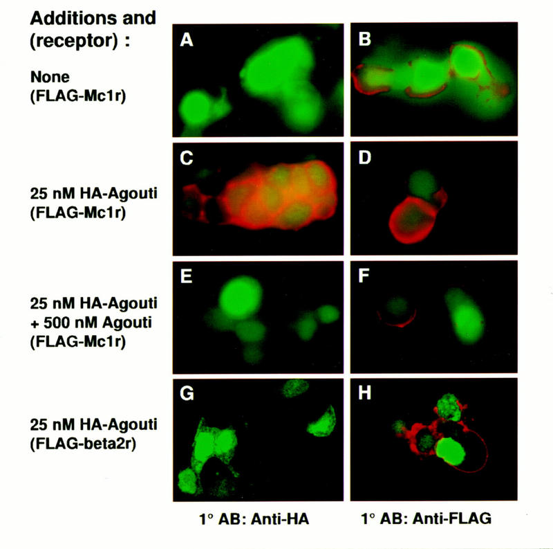
Binding of epitope-tagged Agouti protein to Mc1r-expressing cells. 293T cells were cotransfected with expression plasmids for GFP and the Flag–Mc1r (A–F) or the Flag–B2AR (G,H) as described in Materials and Methods. Transfected cells were incubated in 0 nm Agouti (A,B), 25 nm HA–Agouti (C,D,G,H), or 25 nm HA–Agouti plus 500 nm (untagged) Agouti protein (E,F). Binding of HA–Agouti was detected by immunofluorescence with an anti-HA antibody (A,C,E,G); expression of the Flag–Mc1r or the Flag–B2AR was detected by immunofluorescence with an anti-Flag antibody (B,D,F,H) as described in Materials and Methods. Green staining represents GFP expression and red staining represents bound HA–Agouti (A,C,E,G) or receptor expression (B,D,F,H). All panels represent experiments summarized in Table 1.