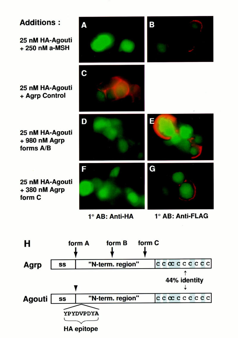
Figure 5.

Effects of Agrp and α-MSH on Agouti protein binding. 293T cells expressing GFP and the Flag–Mc1r were incubated at 4°C in 25 nm HA–Agouti with the following additions: 250 nm α-MSH (A,B), 980 nm Agrp forms A+B (D,E), 380 nm Agrp form C (F,G), or control protein for Agrp purification (C). Binding of HA–Agouti or expression of the Flag–Mc1r was detected by immunofluorescence with anti-HA (A,C,D,F) or anti-Flag (B,E,G) antibodies as described in Materials and Methods. Green staining represents GFP expression; red staining represents bound HA–Agouti (A,C,E,G) or receptor expression (B,E,G). All panels represent experiments summarized in Table 1. (H) Diagram of Agrp and Agouti protein indicating the signal sequence (ss), placement of the HA epitope in HA–Agouti, the different forms of Agrp, and amino acid similarity between Agrp and Agouti, which is confined entirely to the cysteine-rich carboxyl terminus.
