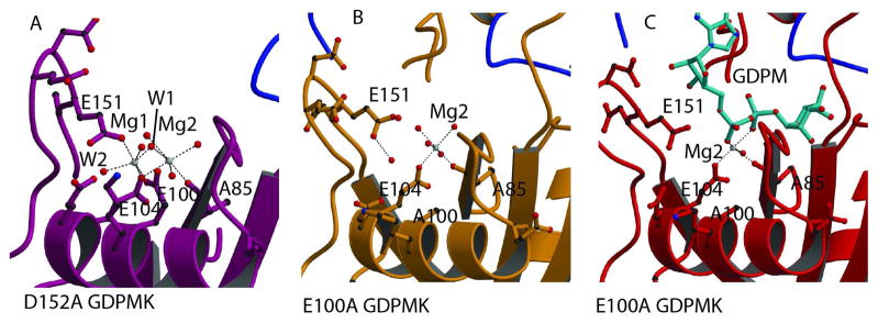
Fig. 2. Mg2+ binding sites.
A. Magnesium binding sites in the structure of the GDPMK D152A mutant. The two magnesium ions are bound to residues of the Nudix signature sequence. Mg1 coordination is completed by E151 from loop L9. B. Metal binding site in the structure of the GDPMK E100A mutant in the absence of substrate. C. Metal binding site in the structure of the GDPMK E100A mutant in the presence of substrate.

