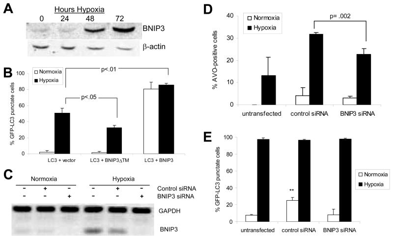
Figure 6.
BNIP3 plays a role in hypoxia-induced autophagic cell death. (A) Western blot showing upregulation of BNIP3 in hypoxia in HEK293 cells. (B) HEK293 cells transfected as in Fig. 6A were incubated in normoxia or hypoxia for 24-hours post-transfection. GFP-LC3 distribution (in co-transfected cells only) was assessed as in Fig. 1B. (C) U87 cells were untransfected, or transfected with control siRNA or BNIP3 siRNA and silencing of gene expression was verified by RT-PCR analysis as described in Materials and Methods. (D) Two days after siRNA silencing, U87 cells were retained in normoxia or exposed to 24 hours hypoxia, and AVO production was measured as in Fig. 1E. (E) Two days after siRNA silencing, U87-GFP-LC3 cells were retained in normoxia or exposed to 24 hours hypoxia, and GFP-LC3 distribution was assessed and quantified as in Fig. 1C. Error bars indicate standard deviation of three independent experiments. Statistical significance was determined by an unpaired t-test: **p<.01.

