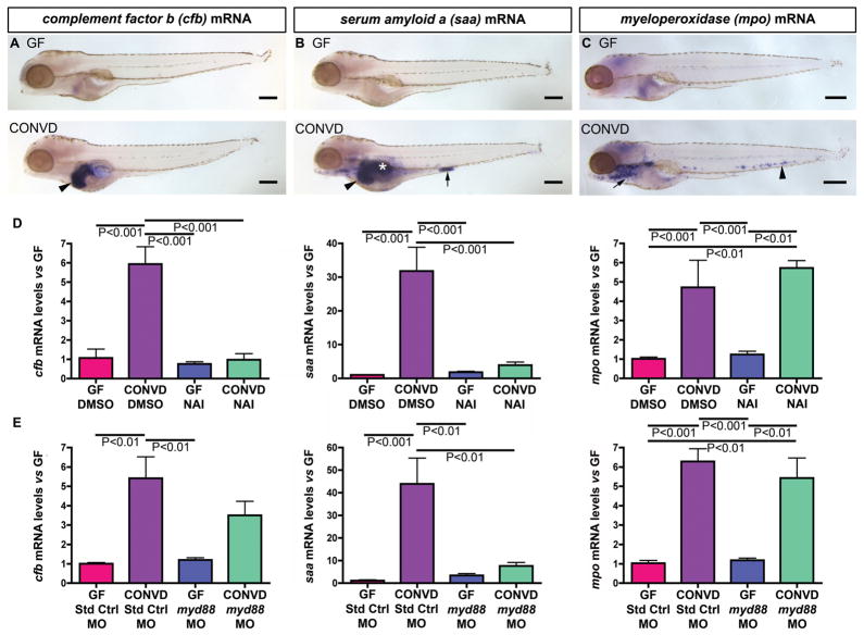
Fig. 6. Conventionalization of GF zebrafish stimulates NF-κB dependent immune responses in diverse cell types.
(A–C) Semi-quantitative wholemount in situ hybridization of 6dpf GF and CONVD zebrafish using RNA probes for complement factor b (cfb), serum amyloid a (saa), and myeloperoxidase (mpo). (A) CONVD zebrafish show increased cfb expression in the liver (black arrowhead) compared to GF controls. (B) CONVD zebrafish also display increased saa expression in liver (black arrowhead), swim bladder (white asterisk) and segment 3 of the intestine (black arrow) compared to GF controls. (C) CONVD larvae robustly express mpo in neutrophils in the caudal hematopoietic tissue (black arrowhead) and other locations (black arrow) compared to GF zebrafish. (D) qRT-PCR assays of whole 6dpf GF and CONVD zebrafish treated with DMSO vehicle from 3–6dpf shows that cfb, saa, and mpo are all significantly induced by the microbiota, and that treatment with 200nM NAI from 3–6dpf attenuates microbial induction of cfb and saa, but not mpo. (E) qRT-PCR assays of whole 6dpf GF and CONVD larvae injected at the 1-cell stage with either standard control morpholino (Std Ctrl MO) or a morpholino targeting myd88 (myd88 MO). qRT-PCR data in panels D–G are from biological duplicate pools (5–20 fish/pool) normalized to 18S rRNA levels and expressed as mean mRNA fold-difference ± SEM. Scale bars: 200μm (A–C).

