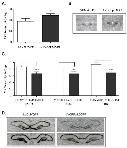
Figure 4. PVN AVP transcript is increased and Hippocampal MR transcript is decreased following CeA CRF-OE (Experiment-1).
Chronic overexpression of CeA CRF increases expression of AVP transcript (nCi/g) in the hypothalamic paraventricular nucleus. (A) Data are displayed as mean +/− SEM; p-values reflect results of one-tailed T-test analysis based on the a-priori hypothesis that increased CeA CRF expression would increase expression of AVP in the (B) Representative example of oligo in situ hybridization for AVP. MR transcript expression (nCi/g) is decreased in the hippocampus of rats overexpressing CeA CRF; upper panel images are anterior relative to lower panel images (C) Data are displayed as mean +/− SEM; p-values reflect results of two-tailed T-test analysis. (D) Representative example of riboprobe in situ hybridization for MR transcript. (* p < 0.05; ** p < 0.01; *** p< 0.001).

