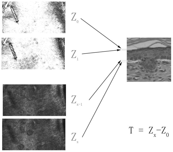
Figure 2. Thickness measurement of viable epidermis from CSLM image analysis.
Sequential stack images from SC to dermis were analyzed. Z0 is defined as the deepest layer where keratinocytes have not yet appeared, Z1 is the layer where keratinocytes begin to appear, Zx−1 is defined as the last layer where basal cells are observed and Zx is defined as the layer where the basal cells are no longer observed. The difference in depth between Zx and Z0 is defined as the thickness of viable epidermis (T).

