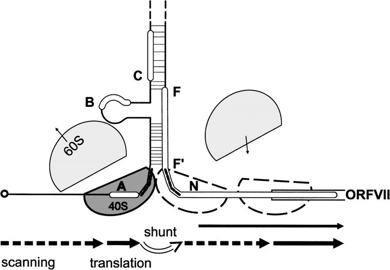
Figure 8.
Model for ribosome shunt. Schematic presentation of the secondary structure of the part of the 35S RNA leader with ORF VII is shown. Broken lines on the structure indicate the positions of the shunt takeoff and landing sites. Arrows show migration of ribosomes (40S and 60S subunits are shown in grey, with outlines representing the subsequent path of the same 40S subunit) by scanning (broken), translation (black), shunting (white). The small arrows show dissociation and association of 60S. Scanning ribosomes enter the 35S RNA at the capped 5′ end and scan until they reach the sORF A start codon. sORF A is translated and properly terminated by most of these ribosomes. The translation event at sORF A provides a specially modified shunt- and reinitiation-competent ribosome that bypasses stem section 1 and is ready to reinitiate just downstream of the shunt landing site. Reinitiation can occur at non-AUG codons (N), but the majority of postshunt ribosomes reinitiate at the AUG of ORF VII. The positive effect of TAV on ribosomal shunt strongly supports the reinitiation model for shunting in CaMV. Note that the translation event at sORF A also leads to partial melting of stem section 1, allowing some post-translation ribosomes to penetrate into stem section 1 and take the alternative route of linear scanning through the leader.

