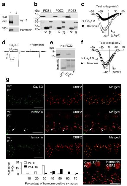
Figure 1.
Harmonin inhibits Cav1.3 channels and is localized at inner hair cell synapses.
(a) Myc-harmonin coimmunoprecipitates with FLAG-α11.3 in cotransfected HEK293T cells (lane 1) but not in cells transfected with myc-harmonin alone (lane 2). (b) Western blot (upper panel) from pull-down assay shows GST-1.3CT (CT) but not GST (G) binds his-tagged PDZ2 of harmonin. Input (I) represents ~5% protein His-PDZ protein input. Ponceau staining (lower panel) shows amounts of GST-proteins used. Full length blots are presented in Supplementary Fig.5. (c) Harmonin inhibits Cav1.3 IBa density in transfected HEK293T cells. Cav1.3 alone, n=11; +harmonin, n=11. (d) Representative traces showing gating currents (Igating) of Cav1.3 ± harmonin measured at the IBa reversal potential (+60 mV). (e) His-PDZ2 of harmonin binds to α11.3 CT but not with L-A mutation. Pull-down assay was done as in b. (f) No effect of harmonin on IBa density for Cav1.3 with L-A mutation (Cav1.3L-A). Cav1.3L-A alone, n=8; Cav1.3+harmonin, n=7. (g) Confocal projections of whole mounts of Organ of Corti from wild-type (WT, P7 or P15) or Cav1.3−/− mice (P7) double-labeled for CtBP2 (red) and Cav1.3 (green) or harmonin (green). Synaptic labeling of CtBP2 appears as spots basal to the nucleus, which is also labeled by these antibodies. Areas of colocalization (arrows) are yellow in the merged images. Scale bars, 10 μm. Graph shows distribution of harmonin-positive synapses in individual IHCs from P6–8 and P14–16 IHCs. In c and f, error bars represent s.e.m.

