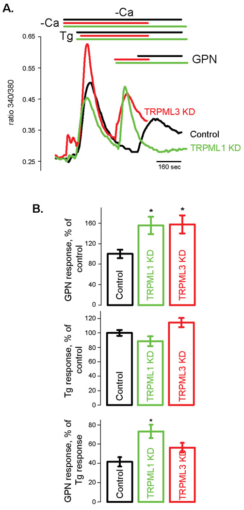
Figure 1. Lysosomal Ca2+ content is increased in TRPML1 deficient HeLa cells.
HeLa cells were transfected with TRPML1 siRNA as described before [79] and cytoplasmic Ca2+ was measured 48 hours later using Fura 2AM. A) Ca2+ traces (cytoplasmic Ca2+ is proportional to the ratio of Fura 2AM fluorescence ratio measured at 340 and 380 nm excitation light). Lysosomes were burst using exracellular application of glycyl-L-phenylalanine 2-naphthylamide (GPN, 100 µM) [44]. Thapsigargin (Tg, 1 µM) was applied before GPN in order to deplete Ca2+ in the endoplasmic reticulum and remove its contribution to the GPN-induced Ca2+ release. Data represent 3–10 experiments and are expressed as mean ± S.E.M. * denotes p<0.05. B) Statistical analysis of Ca2+ measurements. Top: amplitudes of GPN-induced Ca2+ release expressed as a percentage of Ca2+ release in control cells. Middle: Tg-induced Ca2+ release expressed as a percentage of Ca2+ release in control cells. Bottom: the ratios of GPN-induced Ca2+ release to Tg-induced Ca2+ release in control, TRPML1 and TRPML3 deficient cells.

