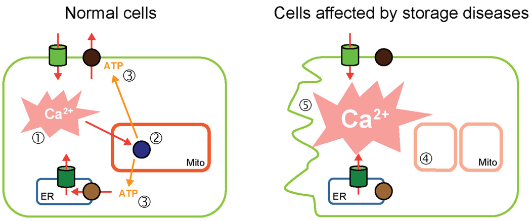
Figure 3. A model depicting mitochondrial deterioration and cell death in storage diseases.
In the normal cells (left) Ca2+ fluxes induced by hormones and neurotransmitters (step 1) are buffered by the energized mitochondria. The Ca2+ dependent components of oxidative phosphorylation chain (step 2) respond by producing more ATP. The resulting spike in ATP promotes Ca2+ extrusion and prevents pro-apoptotic effects of Ca2+. In cells with lysosomal storage diseases (right), the general mitochondrial function is impaired due to buildup of dysfunctional mitochondria (step 4), resulting in the loss of Ca2+/ATP-driven feedback loop, which leads to pro-apoptotic effects of Ca2+ and cell death.

