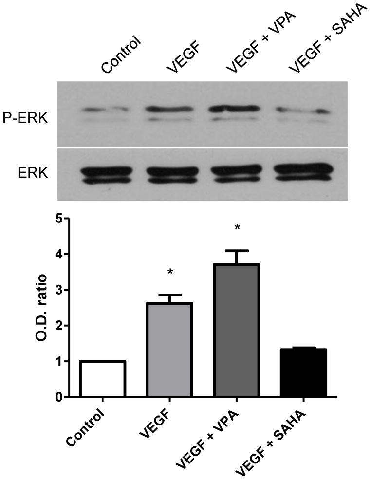
Figure 4.
Western blot for p-ERK and ERK at 6 hours after treatment with VPA or SAHA. Uper panel and bar graph: Presence of VEGF significantly induced p-ERK expression, and the expression of p-ERK greatly increased by addition of VPA. However, the expression of p-ERK was decreased to control level by addition of SAHA. Lower panel: No significant changes were detected in total ERK expressions. * p < 0.05.

