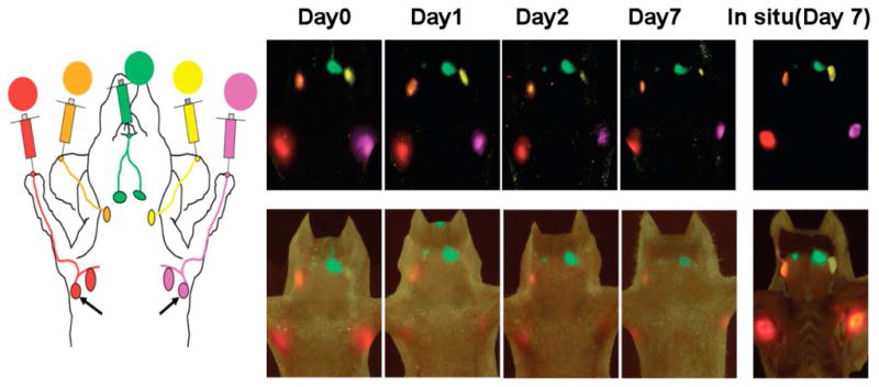
Figure 1.

A five-color optical lymphatic image of lymphatic drainages from the upper extremity (red), the ear (yellow) and the chin (green) obtained using 5 NIR Qdots (Qdots 655,585,545,565, and 605) from day 1 to day 7. There are less marked reductions in signal, except in the left deep neck lymph node (Qdot 565) and a spectral fluorescence imaging technique is shown together with a schematic illustration. (Reproduced from Kosaka et al. [56] with permission)
