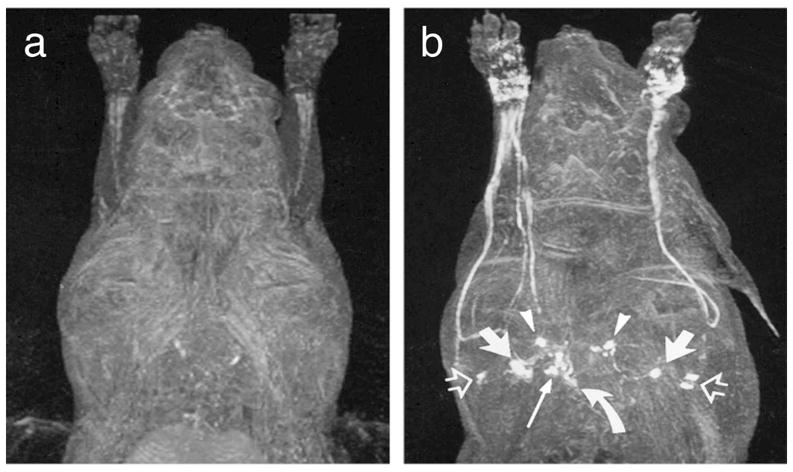
Figure 2.

MRI lymphography demonstrates before (a) and 15 min after (b) subcutaneous administration of gadoterate meglumine bilaterally in the dorsal aspect of the forepaw. The following lymph node groups are depicted in axillary node, anterior thoracic lymph node (arrowheads), parasternal lymph node (curved arrow), and mediastinal lymph node (long solid arrow). (Reproduced from Ruehm et al. [70] with permission)
