
Figure 4.
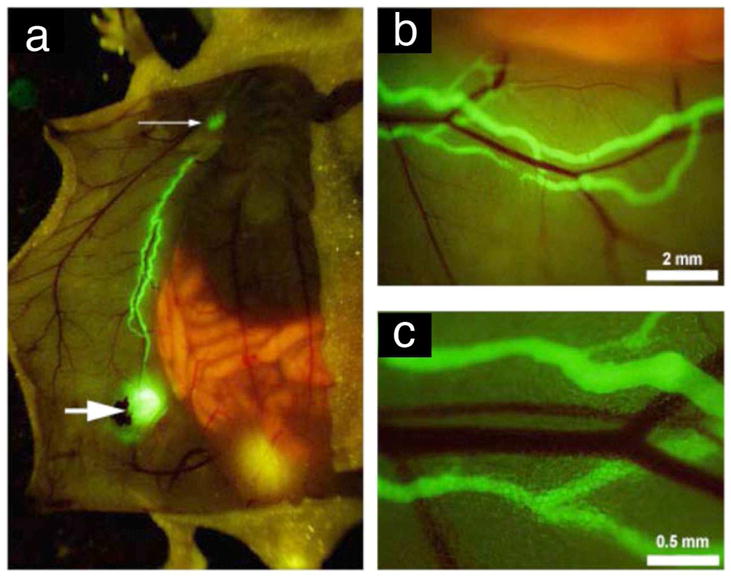
Fluorescence imaging of in vivo of the inguinal lymph nodes (larger arrow), axillary (small arrow) and the connecting lymphatics of the anterior abdominal wall after delivery of AlexaFluor-conjugated monoclonal anti-mouse LYVE-1 antibody (a–c). Neighboring blood vessels did not stain (b,c). (Reproduced from McElroy et al. [151] with permission)