
Figure 2.
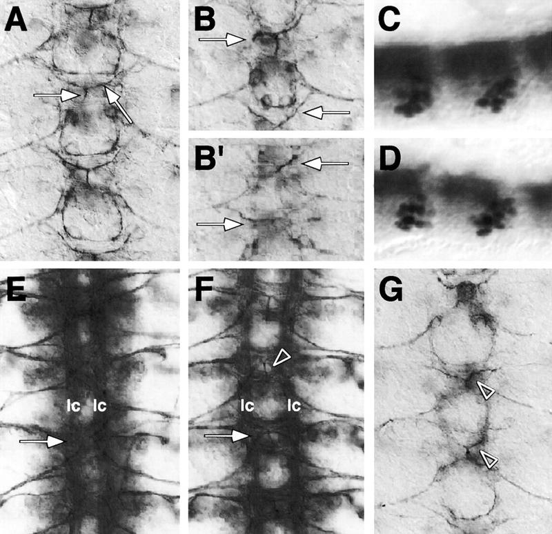
kette functions in midline neurons. Frontal views (A,B,E–G) of dissected embryonic CNS preparations; anterior is up. C and D are whole mount stainings of stage 16 embryos; anterior is to the left. CNS axon tracts are labeled in brown using mAb 22C10. The midline neurons (C,D) are labeled in blue using the enhancer trap insertion X55. (A) In stage 14 wild-type embryos, the VUM axons originate in the midline, project through the posterior commissure to the anterior commissure, where they bifurcate and turn to finally leave the CNS (arrows). (B,B′) In kette mutant embryos, the VUM axons project abnormally. In every neuromere, variable defects in the projection pattern can be observed (arrows, cf. with A). (C) Lateral view of a wild-type stage 16 embryonic CNS. (D) Lateral view of a kette mutant CNS stage 16 embryo. The number and position of the midline neurons has been determined using the marker X55. No changes in number and only slight changes in position were found. (E) In a stage 16 ketteJ4–48 mutant embryo, commissures are fused (arrow) and the two longitudinal connectives (lc) are closer toward the midline. (F) In a stage 16 UAS-kette; simGAL4 mutant ketteJ4–48 embryo, we observe a partial rescue of the kette commissure phenotype (arrow). A space appears between anterior and posterior commissure, and the two longitudinal connectives are located in further distance from the midline. The arrowhead indicates a bifurcating VUM axon. (G) Stage 13 wild-type embryo expressing elevated levels of kette in all midline cells (UAS-kette/simGAL4). Defects in the projection pattern of the VUM neurons can be observed (arrowheads, cf. with A).