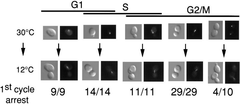
Figure 1.
glc7-129 point of execution is in G2/M. Images of glc7-129 NUF2-GFP (YAB128) cells logarithmically growing at 30°C were captured using DIC (left) and fluorescence (right) microscopy. The fluorescence images represent localization of the spindle pole body(s) (as detected by a Nuf2p–GFP fusion). After shift to 12°C for ∼20 hr, images were captured of the same cells. The numbers below each panel indicate the number of cells that arrest in the first cell cycle as large-budded cells with a short spindle.

