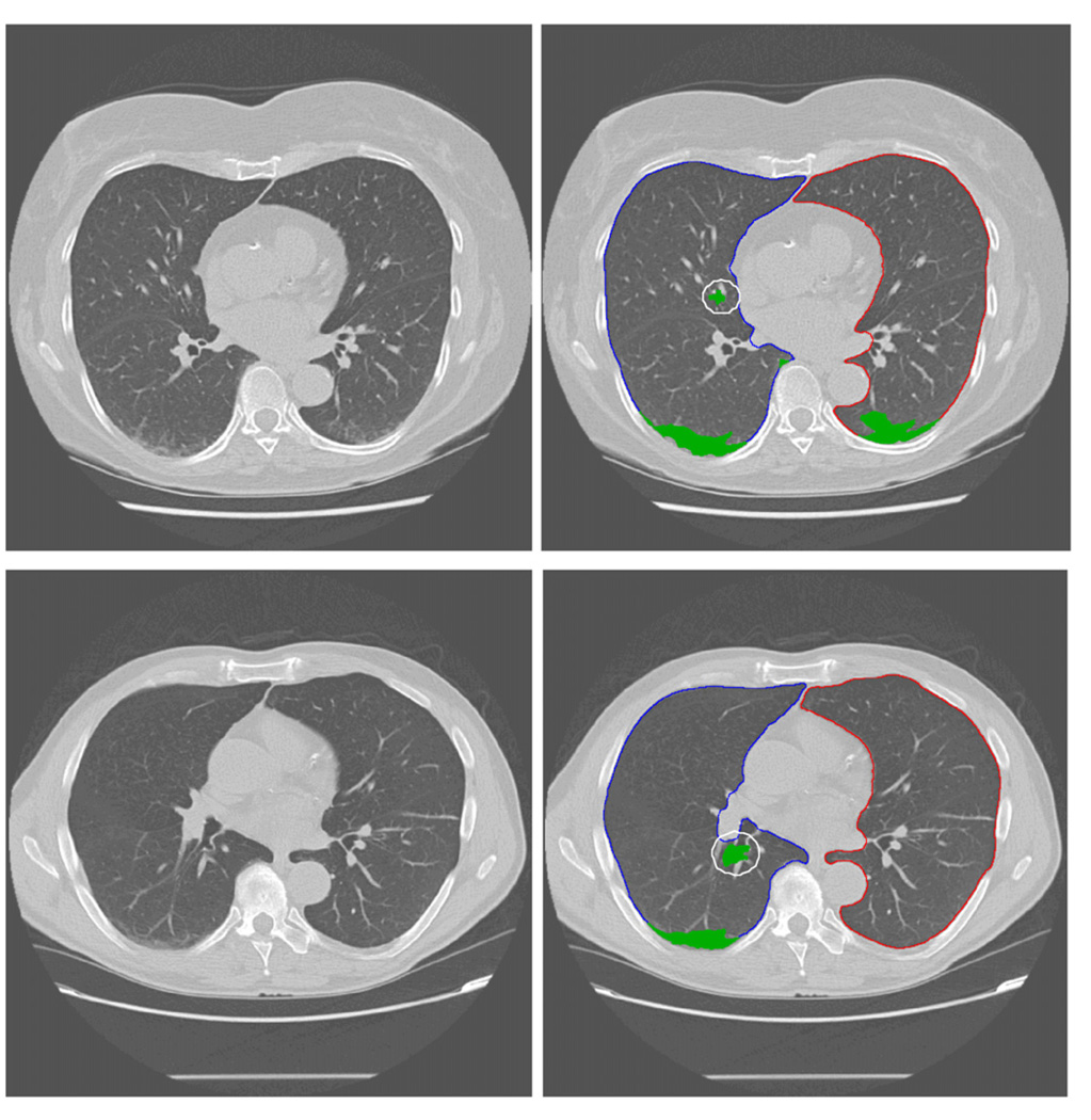
Figure 2.
Examples of partial detection results using the CAD scheme. Two images in the first column and images in the second column are the original images selected from different CT scans for demonstration and the images marked with CAD-generated results (including the boundary contour of the segmented lung areas and identified ILD lesions), respectively. The white circles in the result images indicate false positives because the tissue around vessel is similar to the ILD texture pattern.

